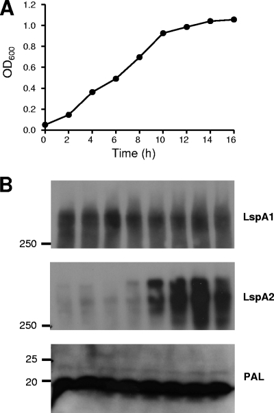
FIG. 1.
Expression of LspA1 and LspA2 during growth of H. ducreyi in broth. (A) Growth of wild-type H. ducreyi 35000HP in CB. (B) Western blot-based detection of LspA1 and LspA2 proteins in whole-cell lysates using the LspA1-specific MAb 40A4 (top panel), the LspA2-specific MAb 1H9 (middle panel), and the PAL-specific MAb 3B9 (bottom panel). This latter antigen was used as a loading control. Cells were sampled every 2 h, beginning with the 2-h time point. Molecular mass position markers (in kilodaltons) are present on the left sides of these three panels.

