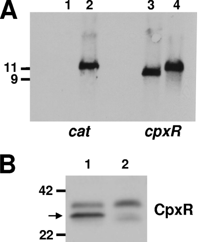
FIG. 4.
Characterization of the H. ducreyi 35000HP cpxR deletion mutant. (A) Southern blot analysis of EcoRV-digested chromosomal DNA from 35000HP (lanes 1 and 3) and the 35000HPΔcpxR mutant (lanes 2 and 4) probed with a cat gene fragment (lanes 1 and 2) and with a cpxR gene fragment (lanes 3 and 4). Size markers (in kb) are present on the left side of this panel. (B) Western blot analysis of whole-cell lysates from 35000HP (lane 1) and the 35000HPΔcpxR mutant (lane 2) probed with polyclonal antibody to the H. ducreyi CpxR protein. The arrow indicates the position of the CpxR protein. Molecular mass position markers (in kilodaltons) are present on the left side of this panel.

