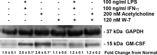
FIG. 7.
Amø GM-CSF protein levels after calmodulin inhibition. Amø from normal rats were incubated with saline, IFN-γ, LPS, or acetylcholine (Ach) for 2 h at the concentrations stated. Some samples were also treated with the calmodulin inhibitor W-7. Soluble Amø proteins were probed for GAPDH and GM-CSF detection. The change in GM-CSF signal strength, normalized to GAPDH signal, was calculated from the average ± SD. Signal strength values were from three individual trials; each trial consisted of Amø pooled from four rats. *, P < 0.05 versus Amø incubated with saline.

