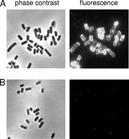
FIG. 3.
Immunofluorescence microscopy. Log-phase cultures of 98NK2sab::kan(pBsab) (A) and 98NK2sab::kan(pB) (B) were formalin fixed and labeled with anti-Sab followed by Alexa 594-conjugated donkey anti-mouse secondary antibody (see Materials and Methods). The fluorescent image for each strain is accompanied by the phase-contrast image for the corresponding field.

