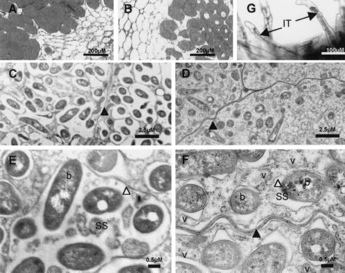
Figure 3.
Microscopic analysis of nodules and infection threads formed by R. etli wild-type (A, C, and E) and casA mutant FAJ1805 (B, D, F, and G) strains. (A and B) Toluidine blue stainings of 3-μm-thick sections of P. vulgaris nodules. (C–F) Transmission electron micrographs of 3-week-old nodules. (G) GusA staining of FAJ1806 bacteria expressing a casA–gusA fusion inside the infection threads. Black arrowhead, plant plasma membrane; white arrowhead, symbiosome membrane; black arrow, infection thread (IT); b, bacteroid; SS, symbiosome space; v, vesicle.

