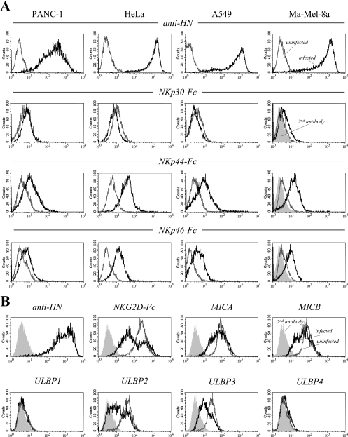
FIG. 1.
Soluble NKp44 and NKp46 receptors bind to various NDV-infected tumor cell lines. (A) PANC-1 pancreatic carcinoma, HeLa cervical carcinoma, A549 lung carcinoma, and Ma-Mel-8a melanoma cells were infected for 20 h with the nonlytic NDV strain Ulster (100 HU/106 cells) (black lines) or left uninfected (gray lines). Cells were stained with NKp30-, NKp44-, and NKp46-IgG1 Fc fusion proteins in complexes with goat anti-hIgG-PE secondary antibodies as indicated. Results for Ma-Mel-8a cells stained with a secondary antibody alone are represented by filled curves. To monitor infection efficiencies, tumor cells were stained with anti-NDV MAb HN.B, recognizing HN (top panels, black lines). For a control, uninfected cells were stained with HN.B (top panels, gray lines). (B) HeLa cells were infected for 18 h with the NDV strain Ulster (black lines) or left uninfected (gray lines). Cells were stained with the NKG2D-Fc fusion protein or with MAbs recognizing NDV HN, to control for infection, or the NKG2D ligands MICA, MICB, and ULBP1 to ULBP4, as indicated. Results for secondary antibody controls are represented by filled curves.

