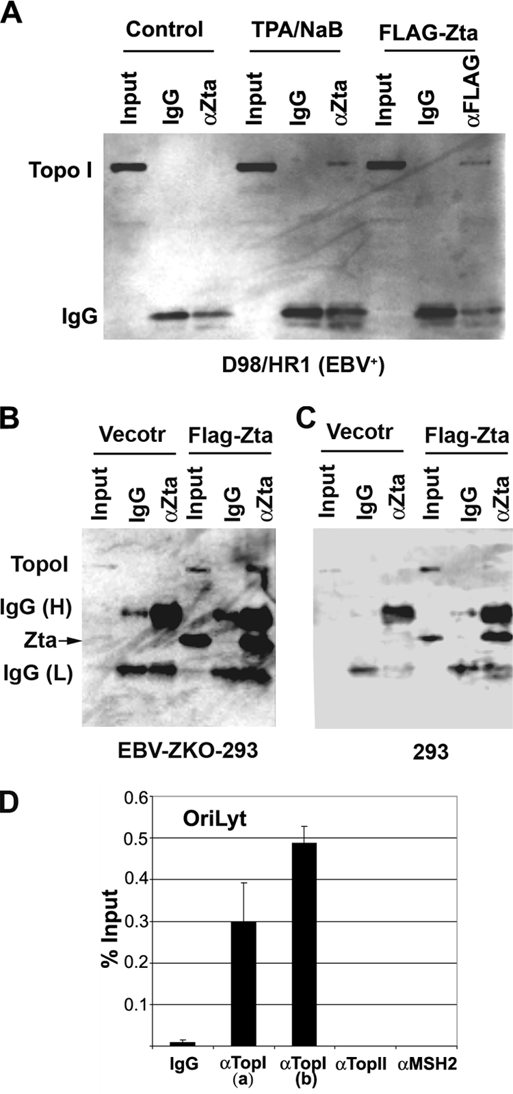
FIG. 4.

Topo I coimmunoprecipitates with Zta and colocalizes with OriLyt. (A) EBV-positive D98/HR1 cells were untreated (lanes 1 to 3), treated with TPA (40 ng/ml) and NaB (3 mM) to induce Zta and lytic replication (lanes 4 to 6), or transfected with FLAG-Zta expression vector (lanes 7 to 9). Cells were then assayed at 48 h posttreatment by immunoprecipitation with antibodies specific for Zta (αZta) (lanes 3 and 6), FLAG (lane 9), or control antibody (IgG) (lanes 2, 4, and 8). Input lanes represent 10% of the starting lysate for each immunoprecipitation. Immunoprecipitates were assayed by Western blotting with Topo I-specific antisera. (B) Western blotting of immunoprecipitates from EBV-positive ZKO-293 cells after transfection with CMV-FLAG vector or CMV-FLAG-Zta expression plasmids. Input (5%), IgG, and anti-Zta immunoprecipitates are indicated for each set of transfections. Western blots were probed with anti-Topo I-specific antibody and visualized by chemiluminescence.(C) Same as B except that EBV-negative 293 cells were used for transfection. (D) D98/HR1 cells treated with TPA and NaB as described above (A) were assayed by ChIP with two different antibodies (a or b) to Topo I, antibodies to Topo II or MSH2, or control IgG, as indicated. ChIP DNA was quantified for EBV OriLyt DNA as a percentage of the total input DNA.
