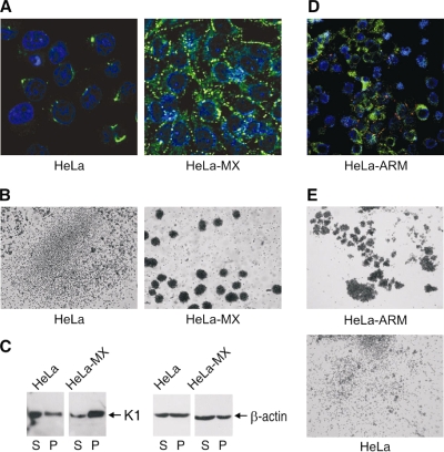
FIG. 3.
Formation of desmosomes in MX LCMV-infected HeLa cells. (A) Immunofluorescence analysis of desmosomes using the specific MAb ZK-31 (green) revealed a lack of desmosomal signal in noninfected HeLa cells. In contrast, HeLa-MX cells showed strong staining signals with a pattern typical for desmosomes. Cell nuclei were stained with DAPI (blue). (B) HeLa-MX cells also exhibited increased cell adhesion capacity in an assay based on spontaneous aggregation of cells first grown in a monolayer and then brought to single-cell suspension, placed on a nonadhesive surface, and rotated overnight on a gyratory shaker. (C) K1 assembly in soluble (S) and pellet (P) fractions of the cell extracts was analyzed by Western blotting. β-Actin was detected as a loading control. (D) Double-staining immunofluorescence analysis of viral NP (green) and desmosomes (red) in HeLa cells persistently infected with LCMV strain Armstrong. Infected cells show a clear desmosomal signal which is absent from noninfected counterparts. (E) An aggregation assay of infected HeLa-ARM cells compared to noninfected control HeLa cells confirmed increased clustering of infected cells.

