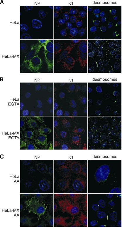
FIG. 4.
Effect of keratin network disassembly on distribution of NP, K1, and desmosomes, showing immunofluorescence analysis of control HeLa and HeLa-MX cells (A) and of cells treated with the inhibitors of intermediate filaments EGTA (B) and acrylamide (AA) (C). The cells were treated for 15 min with 2 mM EGTA and for 8 h with 5 mM AA, and then they were fixed and stained with antibodies specific for NP (green), K1 (red), and desmosomes (green). Cell nuclei were stained with DAPI (blue). Both inhibitors caused a loss or reduction of K1 and desmosomal staining signals in the noninfected cells, whereas these signals were relatively well preserved in the infected cells. Original magnification, ×63.

