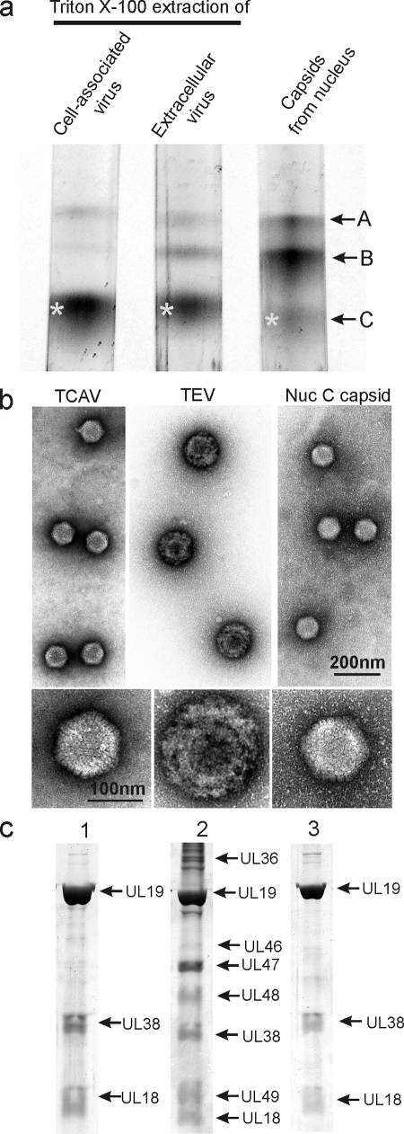
FIG. 2.
Analysis of HSV-1 capsids by sucrose density gradient centrifugation (a), electron microscopy (b), and SDS-PAGE (c). Analysis was performed with capsids isolated by TX-100 treatment of cell-associated virus (lane 1), TX-100 extraction of extracellular virus (lane 2), and capsids isolated from the nuclei of HSV-1-infected cells (lane 3). Bands marked by asterisks in panel a were the ones used for analysis in panels b and c. Note that tegument remains attached to capsids derived from extracellular, but not cell-associated virus.

