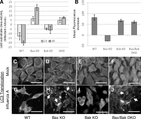
FIG. 6.
Influenza A virus induces autophagy-like death in the absence of Bax. (A) Lysosomal acid phosphatase activity in influenza A virus-infected cells was assessed by measuring the release of p-nitrophenol from p-nitrophenylphosphate substrate. In cells constitutively expressing Bax, acid phosphatase activity decreases following infection, as these cells follow an apoptotic path to death. In cells lacking Bax, acid phosphatase activity increased, indicating an increase in lysosomal activity, a common marker for autophagic death. One unit equals 1 μmol of 4-nitrophenylphosphate per minute under the experimental conditions. (B) Cells were infected for 48 hpi and stained with Lysotracker Red DND-99 for 30 min prior to collection and immediate analysis by FACS. In WT, Bak KO, and Bax/Bak DKO cells, influenza A virus infection results in an increase in lysosomal volume by 48 hpi. In Bax KO cells, a slight decrease in lysosomal volume was observed by 48 hpi. (C to J) Cells were transfected with a construct expressing LC3-GFP prior to infection and observed by confocal microscopy for LC3 expression and translocation following infection. (C to F) The diffuse LC3-GFP expression pattern in mock-infected cells indicates a cytoplasmic, inactive distribution of LC3 in each cell type. (G and I) In cells constitutively expressing Bax, LC3-GFP expression remains diffuse following infection, indicating a lack of LC3 activation as these cells undergo apoptosis. (H and J) In cells lacking Bax (Bax KO and Bax/Bak DKO cells), LC3-GFP staining shifts to a punctate pattern, indicating LC3 activation and translocation to autophagosomes, a process that occurs solely during autophagy. Bars, 25 μm.

