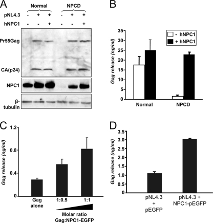
FIG. 6.
Exogenous NPC1 restores Gag release in NPCD cells to normal levels, and NPC1 overexpression correlates with increased Gag release. (A, B) Normal or NPCD fibroblasts were cotransfected with the pNL4.3 proviral clone and either an NPC1 expression vector or empty vector. Samples were harvested after 24 h of transfection. (A) Cell lysates were collected and analyzed by immunoblotting with anti-NPC1 antibody after normalizing by amount of protein (top blot). The level of Gag protein and β-tubulin was detected by Western blotting with anti-Gag (middle) and anti-β-tubulin (bottom). (B) Amount of Gag released in the supernatant was quantified by a p24 ELISA. Data shown represent the mean ± standard deviation from two independent experiments. (C) Overexpression of NPC1 in TZM-bl cells increases Gag particle release. TZM-bl cells were transfected with a Gag plasmid and with increasing amounts of NPC1-enhanced GFP DNA, as indicated. Gag released in the supernatant was quantified by p24 capture ELISA. Data shown represent the mean±SD from two independent experiments. (D) Overexpression of NPC1 in TZM-bl cells increases viral particle release. TZM-bl cells were transfected with pNL4.3 proviral DNA and cotransfected with either pEGFP or NPC1-enhanced GFP. The amount of Gag released in the supernatant was quantified 48 h post transfection using p24 ELISA. Data shown represent the mean ±SD from two independent experiments.

