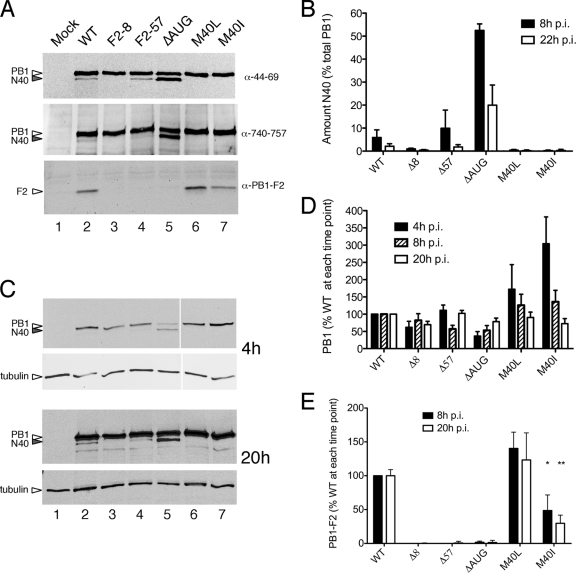
FIG. 2.
Expression of segment 2 polypeptides in virus-infected cells. MDCK cells were infected (or mock infected) with the panel of viruses as labeled, and cell lysates were obtained at various times p.i. (panel A, 8 h p.i.; panels C to E, as labeled) analyzed by Western blotting for the indicated polypeptides. Representative experiments are shown in panels A and C. (B, D, and E) The accumulation of the indicated polypeptides was quantified. The means ± the standard errors of the mean (SEM) of four independent experiments using two independently rescued virus stocks are plotted in panels B and D, while the data in panel E reflect the means and ranges of two independent experiments.

