FIG. 3.
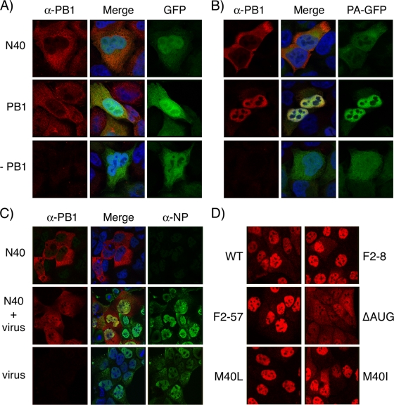
Intracellular localization of PB1-related polypeptides. (A and B) HeLa cells were transfected with plasmids encoding N40, PB1, GFP, or PA-GFP as indicated; fixed 24 h later; stained with anti-PB1 MAb 10.4; and imaged by confocal microscopy. (C) MDCK cells transfected with plasmids encoding N40 or (bottom row) empty vector were infected 24 h later with Dk/Sing virus and at 8 h p.i. were fixed and stained with anti-PB1 MAb 10.4 and anti-NP 2915 before imaging by confocal microscopy. (D) MDCK cells were infected with the indicated viruses, fixed, and stained with anti-PB1 MAb 10.4 at 8 h p.i. Confocal settings were kept consistent throughout each experiment.

