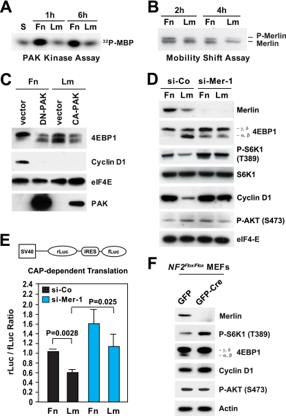
FIG. 3.
Inactivation of merlin mediates integrin-dependent mTORC1 signaling. (A) Integrin-specific adhesion controls activation of PAK. HUVECs synchronized in G0 were detached and kept in suspension (S) or plated onto fibronectin (Fn) or laminin 1 (Lm) in the presence of mitogens for the indicated times. Equal amounts of total proteins were subjected to immunoprecipitation with antibodies to PAK, followed by an in vitro kinase assay using 32P-labeled myelin basic protein (32P-MBP) as a substrate. (B) Integrin-specific adhesion promotes phosphorylation of merlin. HUVECs were transfected with a plasmid encoding wild-type merlin. After synchronization in G0, the cells were plated onto fibronectin or laminin 1 in the presence of mitogens for the indicated times. Equal amounts of proteins were subjected to high-resolution SDS-PAGE, followed by immunoblotting with antimerlin. P-merlin, phosphorylated merlin. (C) PAK activity is necessary for phosphorylation of 4EBP1 and expression of cyclin D1 on fibronectin. HUVECs were transfected with an empty vector or vectors encoding dominant negative (DN) or constitutively active (CA) PAK. After synchronization in G0, the cells were plated onto fibronectin or laminin 1 in the presence of mitogens for 24 h and lysed. Equal amounts of total proteins were subjected to immunoblotting with antibodies to the indicated antigens. (D) Depletion of merlin rescues phosphorylation of 4EBP1 and expression of cyclin D1 on laminin 1. HUVECs were transfected with an siRNA oligonucleotide targeting human merlin (si-Mer-1) or control siRNA (si-Co), synchronized in G0, detached, and plated onto fibronectin or laminin 1 in the presence of mitogens for 24 h. Total lysates were analyzed by immunoblotting using antibodies to the indicated antigens. P-S6K1 (T389), S6K1 phosphorylated at T389; P-AKT (S473), AKT phosphorylated at S473. (E) Depletion of merlin promotes cap-dependent translation. HUVECs were transfected with the bicistronic reporter construct depicted above the graph, in combination with an siRNA oligonucleotide targeting human merlin or control siRNA. Cells were synchronized in G0 and plated onto fibronectin or laminin 1 in the presence of growth factors for 24 h. The graph shows the mean ratios ± SD between the Renilla luciferase (rLuc) and firefly luciferase (fLuc) bioluminescence levels in the indicated samples. SV40, simian virus 40; IRES, internal ribosome entry site. (F) Genetic inactivation of NF2 promotes mTORC1 signaling without activating AKT in primary fibroblasts. NF2flox/flox MEFs were infected with adenoviruses encoding green fluorescent protein-Cre (GFP-Cre) or GFP alone. Two days later, cells were synchronized in G0, plated onto fibronectin for 20 h in the presence of 20 ng/ml bFGF-1 μg/ml heparin, and subjected to immunoblotting using antibodies to the indicated antigens.

