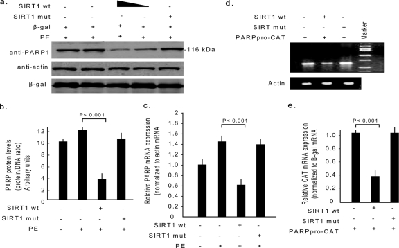
FIG. 6.
SIRT1 negatively regulates PARP1 gene expression. (a) Cardiomyocytes were overexpressed with the SIRT1 wild type (10 and 20 MOI) or the mutant (20 MOI) by use of adenovirus vectors and were then stimulated by PE (20 μM) for 48 h. Cells were also infected with β-Gal adenovirus (10 MOI), which served as the negative control. The cell lysate was analyzed by Western blotting with anti-PARP1, antiactin, and β-Gal antibodies. In the same cell lysate, DNA content was also measured. (b) Quantification of PARP1 content in different groups of cardiomyocytes. (c) Endogenous levels of PARP1 mRNA in different groups of cardiomyocytes, measured by real-time PCR. (d) Cos7 cells were cotransfected with the CAT reporter plasmid, in which the CAT gene was driven by the −237 bp upstream promoter region of the PARP1 gene (PARPPro-CAT), and the expression plasmids encoding either wild-type (wt) or mutant (mut) SIRT1. The gel image shows the amplification of CAT transcripts from different groups of cells under identical conditions. The actin transcript was used as a reference control. (e) CAT mRNA levels in different groups of transfections, measured by real-time PCR and normalized to β-Gal mRNA. All values are means ± SE of the results for four to six different experiments. +, present; −, absent.

