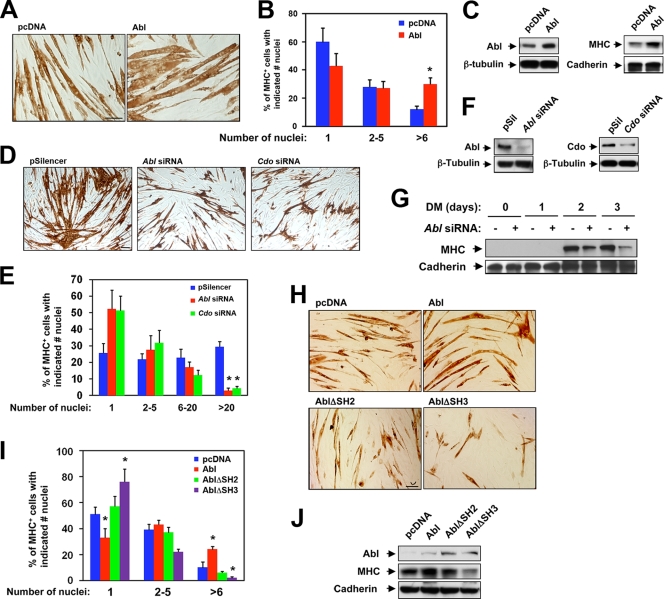
FIG. 4.
Abl promotes myogenic differentiation. (A) Photomicrographs of C2C12 cells that stably express Abl or control (pcDNA) vectors that were cultured in DM, fixed, and stained with an antibody to MHC. (B) Quantification of myotube formation by the cell lines shown in panel A. Values represent means of triplicate determinations ± 1 standard deviation. The experiment was repeated three times with similar results. Asterisks indicate difference from the control at P < 0.01. (C) Lysates of cell lines shown in panel A were Western blotted with antibodies to Abl or MHC and to β-tubulin or pan-cadherin as loading controls. (D) Photomicrographs of C2C12 cells that stably express Abl siRNA, Cdo siRNA, or control (pSilencer) vectors that were cultured in DM, fixed, and stained with an antibody to MHC. (E) Quantification of myotube formation by the cell lines shown in panel D. Values represent means of triplicate determinations ± 1 standard deviation. The experiment was repeated three times with similar results. Asterisks indicate difference from the control at P < 0.01. (F) Lysates of cell lines shown in panel D were Western blotted with antibodies to Abl or Cdo to reveal the level of RNAi-mediated depletion and to pan-cadherin as a loading control. (G) Lysates of control and Abl siRNA cell lines shown in panel D were cultured for 1, 2, or 3 days in DM and then Western blotted with antibodies to MHC and to pan-cadherin as a loading control. (H) Photomicrographs of C2C12 cells that stably express the indicated Abl proteins or a control (pcDNA) vector that were cultured in DM, fixed, and stained with an antibody to MHC. (I) Quantification of myotube formation by the cell lines shown in panel H. Values represent means of triplicate determinations ± 1 standard deviation. The experiment was repeated three times with similar results. Asterisks indicate difference from the control at P < 0.01. (J) Lysates of the cell lines shown in panel H were Western blotted with antibodies to Abl, MHC, and pan-cadherin as a loading control.

