FIG. 2.
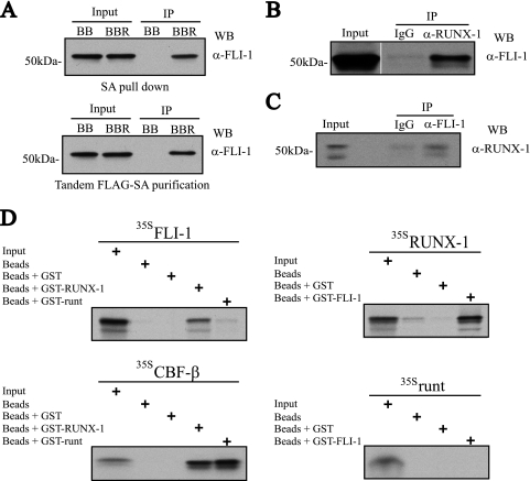
Physical interaction between RUNX-1 and FLI-1. (A) FLI-1 Western blot (WB) analysis of single SA (top)- or tandem anti-FLAG-SA (bottom)-purified FLAG-BioRUNX-1 complexes from TPA-induced L8057 cells. BB, birA plus empty FLAG-biotinylation vector; BBR, birA plus FLAG-BioRUNX-1 expression vector; α-, anti-; IP, immunoprecipitation. (B) Co-IP of endogenous RUNX-1 and FLI-1 from TPA-induced L8057 cells. Immunoprecipitation with normal rabbit IgG or anti-RUNX-1 antibody, and Western blot with anti-FLI-1 antibody. A 2% input is shown. (C) Immunoprecipitation with normal rabbit IgG or anti-FLI-1 antibody, and Western blot with anti-RUNX-1 antibody. A 2% input is shown. (D) Direct RUNX-1-FLI-1 protein-protein interactions. In vitro transcribed and translated [35S]methionine-labeled (35S) FLI-1, RUNX-1, runt domain, or CBF-β was incubated with uncoupled Sepharose beads or beads coupled with bacterially produced GST, GST-RUNX-1, GST-FLI-1, or GST-runt domain, as indicated. The beads were washed, and eluted material was separated by SDS-PAGE. An autoradiogram of the gel is shown. Ten percent of the 35S-labeled input protein is included.

