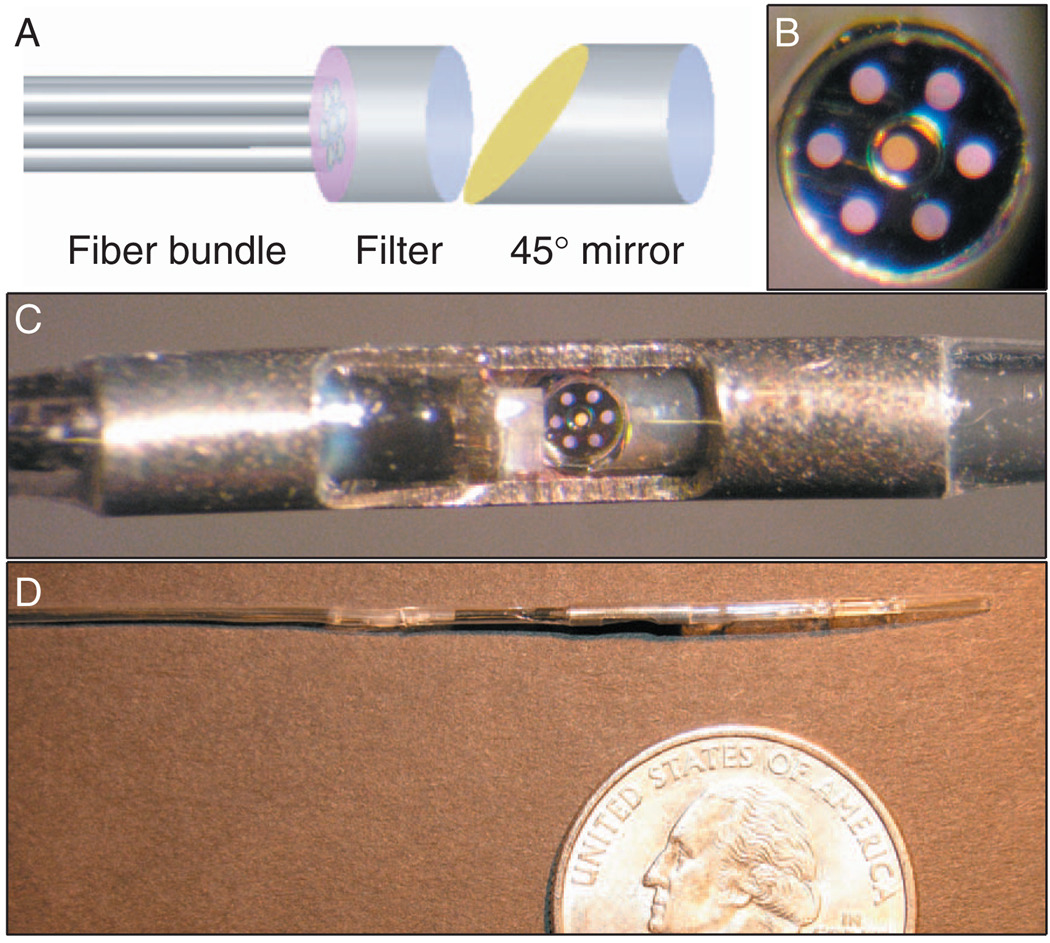
Fig. 1.
(a) Schematic of the Raman catheter distal optics. (b) En face photograph of white light transmission through the fiber bundle and custom filter. The inner fiber (orange) was used for excitation; the six outer fibers (purple) were used for collection. (c) Close-up view of distal end of the Raman catheter. The mirror shows the reflection from the fiber bundle face. (d) Distal portion of the catheter.

