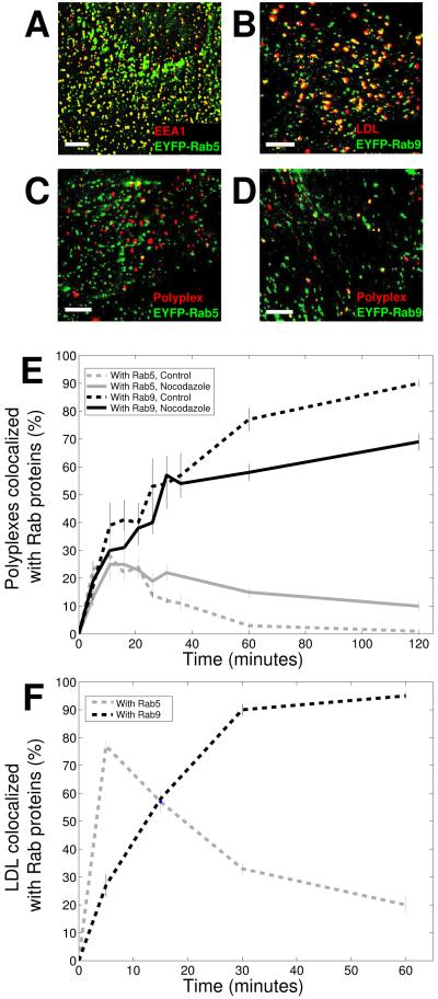
Figure 6. Proteoglycans and proteoglycan-binding ligands are trafficked to late endosomes with high efficiency.
A) Colocalization between EYFP-Rab5 (green) and EEA1 (red), an early endosomal marker. EEA1 was detected by immunofluorescence. Image adapted from Lakadamyali, et al. (43). B) Colocalization between EYFP-Rab9 (green) and DiD-labeled LDL (red). The image was taken 1 hour after LDL internalization, at which point LDL accumulates in late endosomes. C) A two-color image of polyplexes and EYFP-Rab5, an early endosomal marker, 20 minutes after the addition of polyplexes to cells. D) A two-color image of polyplexes and EYFP-Rab9, a late endosomal marker, 1 hour after the addition of polyplexes to cells. Colocalization is shown in yellow. Scale bars: 10 μm. E) Fraction of polyplexes colocalized with Rab5 (gray) and Rab9 (black) as a function of time. The solid and dashed lines indicate results from cells treated with nocodazole and untreated cells, respectively. . Error bars indicate standard error. Results were averaged over 5 cells. F) Fraction of LDL particles colocalized with Rab5 (gray) and Rab9 (black) as a function of time in untreated cells. Error bars indicate standard error. Results were averaged over 4 - 7 cells.

