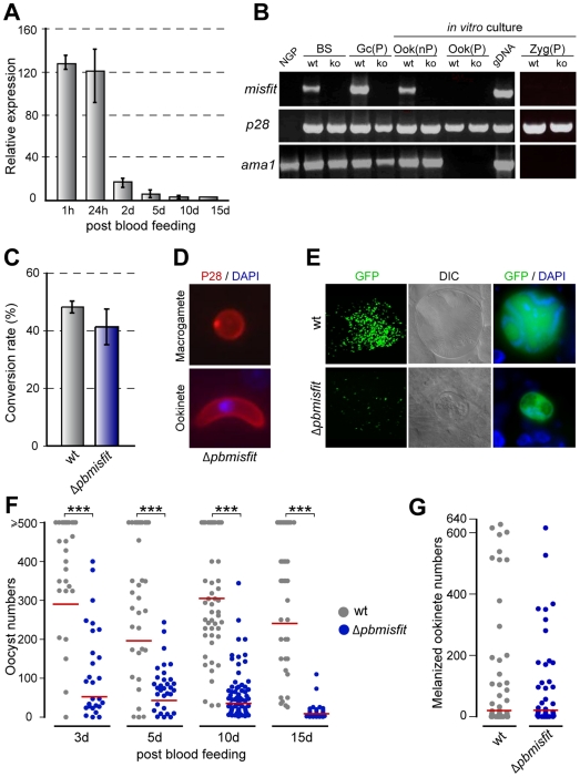
Figure 1. Misfit gene expression and phenotypic analysis of Δpbmisfit mutants.
(A) Relative abundance of misfit transcripts in P. berghei-infected A. gambiae midguts, assayed by quantitative real-time RT-PCR. The constitutively expressed gfp transgene was used as reference. The average expression and standard errors of three independent biological replicates (different batches of mosquitoes fed on different blood sources) are shown. The results of each biological replicate are the average of two technical replicates. (B) RT-PCR analysis of misfit in non-purified (nP) and purified (P) wt and misfit ko parasite populations. Genes encoding the female/zygote sexual stage protein, P28, and the blood-stage protein, Ama1 (apical membrane antigen 1), served as stage-specific and loading controls. NGP, non-gametocyte producing strain; BS, mixed asexual and sexual blood stages; Gc, gametocytes; Ook, ookinetes; gDNA, genomic DNA; Zyg, zygote. (C) Macrogamete to ookinete conversion assay in control wt and Δpbmisfit mutant parasites. (D) In vitro cultured Δpbmisfit parasites stained for P28 (red) and DNA (DAPI, blue). (E) Microscopy images of 15-day-old wt and Δpbmisfit oocysts in A. gambiae midguts (first column ×10 objective; second and third columns ×63 objective). GFP-expressing parasites appear green. DIC, differential interference contrast. (F) Distribution of wt and Δpbmisfit oocyst numbers in midguts of A. stephensi mosquitoes, at day 3, 5, 10 and 15 post blood feeding. The geometric means of oocyst numbers (red line) are shown. Highly significant reduction (1-way ANOVA, P<0.001, ***) of Δpbmisfit oocyst numbers compared to wt controls is detected at all time points. Oocyst numbers equal or higher than 500 were individually enumerated and used to calculate the means. The full analysis is presented in Table S2. (G) Ookinete invasion assay in CTL4 kd A. gambiae. Distribution and geometric means of melanized ookinete intensities are shown. No statistical difference is detected between wt and Δpbmisfit parasites.

