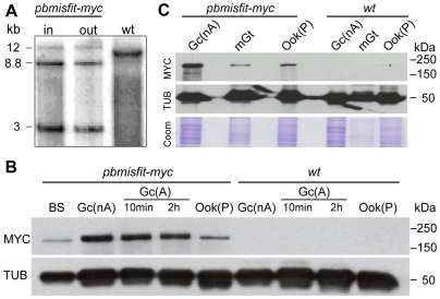
Figure 2. Expression of MYC tagged MISFIT in transgenic P. berghei.
(A) Southern blot analysis of input and output (back-bite) pbmisfit-myc parasite populations demonstrates population purity and stability of the tagged locus that was generated as shown in Figure S3. Genomic DNA was digested with EcoRI and a 3′ UTR fragment of misfit was used as probe. Insertion of the transgenic cassette resulted in a 3 kb digestion product, which is absent from wt parasites. The 8.8 kb band represents a tandem insertion of two tagging vectors in the target misfit locus. The wt band detected for the native misfit locus is absent from pbmisfit-myc populations. (B) Western blot analysis of transgenic pbmisfit-myc parasite populations using an anti-MYC antibody. Wt parasites were used as a control. Tubulin (TUB) detected with a mouse monoclonal antibody against Trypanosoma brucei alpha-tubulin (tat1) was used as a positive control. BS, mixed asexual and sexual blood stages; Gc(nA), non activated gametocytes; Gc(A), activated gametocytes 10 min or 2 h post-activation; Ook(P), purified ookinetes. (C) Western blot analysis of pbmisfit-myc microgametes (mGt) using an anti-MYC antibody. Gc(nA) and Ook(P) extracts were used as a control. TUB was used as internal control, and Coomassie (Coom) stained extracts as a loading control. Numbers indicate protein size scale in kDa.

