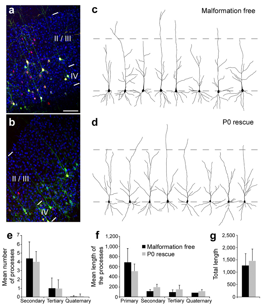
Figure 4. Morphology of rescued neurons.
(a, b) Immunohistochemistry for the upper layers neurons marker CDP/Cux1 on P20 frontal neocortical sections. Animals were electroporated at E14 with either non-effective (3UTRm3hp) (a) or effective (3UTRhp) (b) Dcx-targeting shRNA vectors together with CAG-mRFP, CAG-ERT2CreERT2, and CALNL-eGFP (a) or CALNL-DCX-eGFP (b), and injected with 4-OHT at birth. Both initially correctly positioned transfected neurons (a) and initially mis-positioned transfected neurons induced to migrate to appropriated positions after Dcx re-expression (b) (both green and red) are located within the CDP/Cux1+ band of upper layers neurons (in blue). (c, d) Reconstructed cortical neurons showing dendritic arborization in the same experimental conditions. (e–g) Quantifications of the dendritic arborization: mean number of apical processes per neurons (e), their mean length (f) and total length (g) (36–37 reconstructed neurons from 3–4 animals per condition). Scale bars, 200 µm (a).

