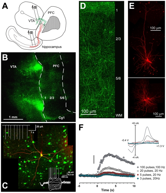
Figure 1. Properties of the organotypic triple co-culture system and the DA innervation of the PFC as demonstrated by tyrosine-hydroxylase (TH) containing fibers.
A) Schematic representation of the triple co-culture consisting of the PFC, VTA, and hippocampus. Electrical stimulation of the afferents from the VTA (indicated as green lines) or ventral hippocampus (red lines) induces Up-states in the PFC. B-D) Photomicrographs illustrating putative DAergic (TH-positive) neurons in the VTA and the distribution of TH-fibers in the PFC. Co-cultures were made from mice expressing green fluorescent protein under the control of the TH gene promoter. C) Properties of putative DAergic (green TH-positive) neurons in the VTA. Cell-attached recordings (top left inset) show that DA neurons are tonically active. Bottom right inset: Membrane properties and firing response in whole-cell mode in response to a series of hyperpolarizing and depolarizing current steps (−150 to+120 pA). The recorded cell was filled with Alexa 594 after break-in. D) shows the laminar distribution of fibers in the PFC. E) Morphological properties of a pyramidal cell (top) and interneuron in the PFC of organotypic co-cultures. Cells were loaded with Alexa 594 during recording and visualized using series of confocal images. Images are montages of convoluted z-stacked images at 40× magnification in C-F. All images were contrast-enhanced for clarity. F) Electrochemical detection of phasic DA release in the PFC following stimulation of the VTA. Stimulation trains (3–100 pulses) were initiated at time 0, and evoked an increase in extracellular DA. Scale bar is 200 nM. The insert shows background-subtracted cyclical voltammograms taken at the peak of the response for each of the stimulations. Abbreviations: VTA, ventral tegmental area; PFC, prefrontal cortex, Cg1, cingulate cortex; WM, white matter.

