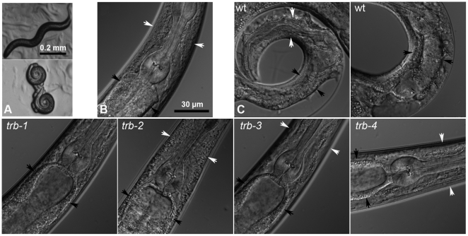
Figure 1. Intoxication of C. elegans by tribendimidine.
A. L4 worms exposed to no drug (upper) or 100 µg/mL tribendimidine (lower) for 24 h at 25° and photographed at 30× magnification. Tribendimidine causes most C. elegans animals to coil. Scale bar applies to both panels. B and C. 600× magnification of animals under various conditions. B. Wild-type control animal without drug showing healthy intestine (between black arrowheads). White arrowheads (here and in other panels) point to cuticular regions within which the pharyngeal isthmus is contained. C. Animals on 100 µg/mL tribendimidine. Top row: wild-type animals on tribendimidine. Significant damage to the intestine (between black arrowheads) is evident, as well as degradation of the body around the pharyngeal isthmus of the left-most animal. Bottom row: tribendimidine resistant animals on tribendimidine. Note, all have healthy intestines and no degradation of body cavity is evident. Scale bar in B applies to all images in B and C. wt = wild type. Alleles used are trb-1(ye492), trb-2(ye493), trb-3(ye494), and trb-4(ye495).

