
Fig. 12.
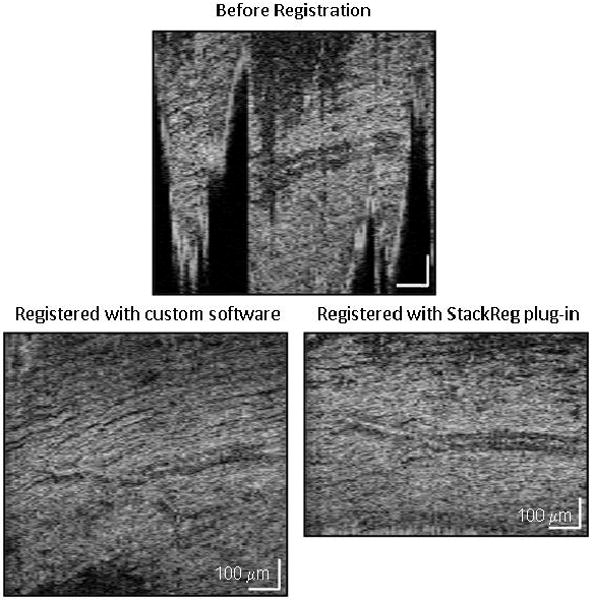
Performance of volume registration software using the “same” C-scan extracted from Case 7: (top) unregistered, (left) registered with custom software, and (right) registered using the “StackReg” plug-in for ImageJ. The C-scan slices through the nerve fiber layer. The volume was acquired at 6 degrees superior of the fovea with focus at the nerve fiber layer. Note the dark regions in the unregistered C-scan correspond to the vitreous and are due to axial motion of the eye during the volume acquisition. White scale bars indicate a length of 100 μm.