
Fig. 7.
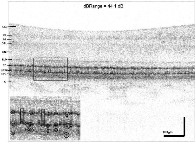
B-scan image extracted from the BroadLighter volume in Fig. 5 (Case 1). Data was encoded over 44 dB above the noise floor and displayed using an inverted gray scale. Note the uneven appearance of the CC that is particularly evident in the 2× magnified view (inset), which shows a highlighted rectangular region at the depth of the photoreceptors. Multiple reflections sometimes occur in the same outer segment (denoted by rectangles in the inset). Also in the inset, the strongest reflections from within the RPE often coincide with reflections from an overlying cone (denoted by circles in the inset). GCL: ganglion cell layer; IPL: inner plexiform layer; INL: inner nuclear layer; OPL: outer plexiform layer; ONL: outer nuclear layer; ELM: external limiting membrane; CC: connecting cilia; OTPR: outer tips of the photoreceptors; RPE: retinal pigment epithelium; C: choroid.