
Fig. 9.
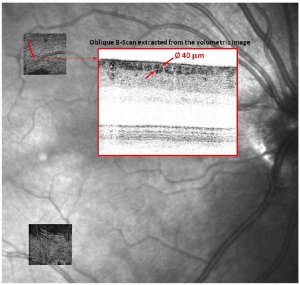
C-scans of Case 5 and Case 7 superimposed on a commercial SLO fundus image taken from the same 29-year old female subject. Both C-scans slice through the RNFL and show large blood vessels that lie at the same proximal depth. (inset) B-scan is an average of two adjacent oblique B-scans extracted from the Case 7 volume. An individual nerve fiber bundle is highlighted whose diameter was measured at 40 μm. Other bundles of similar size are also present.