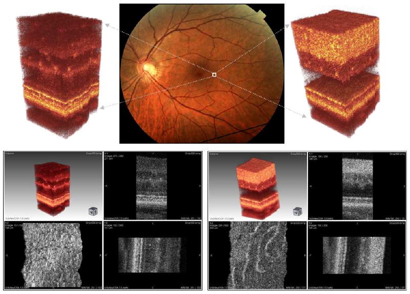
Fig. 5.

UHR-AO-OCT volume of the retinal structures acquired over 0.25 × 0.3 mm area on the 4.5 deg temporal retina (TR). Top row: left, visualization of the data acquired with UHR-AO-OCT system focus set on photoreceptor layers; center, fundus photo with marked location of the acquired volumes; right, visualization of the data acquired with UHR-AO-OCT system focus set on inner retinal layers. Bottom row: left, screenshot from OSA ISP with the UHR-AO-OCT volume with focus set on photoreceptor layers (View 1); right, screenshot from OSA ISP with the UHR-AO-OCT volume and focus set on inner retina layers (View 2). Multiple microscopic and cellular structures can be recognized clearly (including photoreceptors seen on the lower left panel and microcapillaries seen on the lower right panel).
