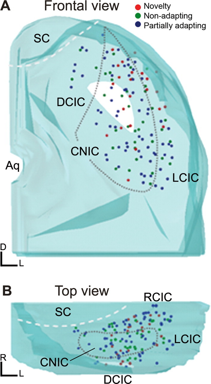
Figure 10.

A, B, Computer-assisted three-dimensional reconstructions of the locations of neurons that were histologically localized, seen in a frontal projection (A) and in a horizontal projection (B), rotated 90° from the projection seen in A. Neurons showing the entire range of SSA were found in all subdivisions of the IC, although a higher proportion of the neurons with the highest degree of SSA were located in the dorsal and rostral parts of the IC. Aq, Aqueduct; DCIC, dorsal cortex of the inferior colliculus; LCIC, lateral cortex of the inferior colliculus; RCIC, rostral cortex of the inferior colliculus; SC, superior colliculus.
