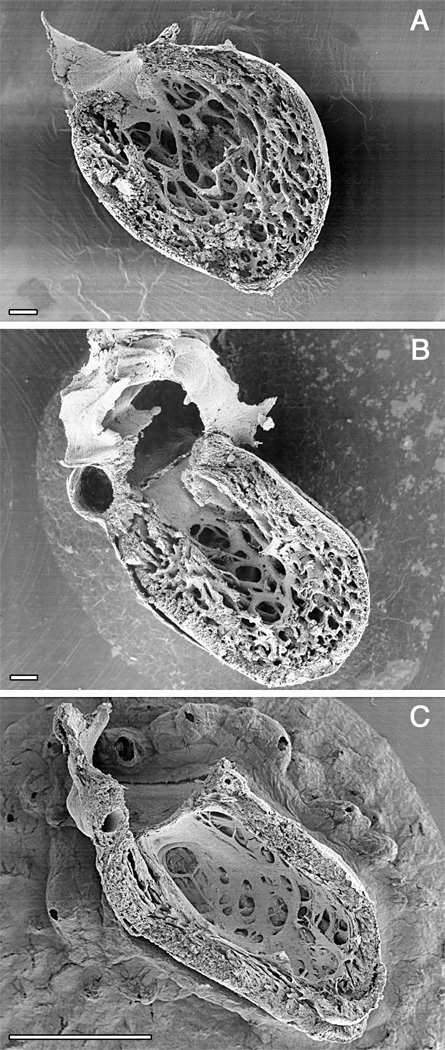
Figure 2.
Time course of ventricular compaction in the human left ventricle. Note increasing proportion and thickness of the outer compact layer. A. Numerous fine trabeculae are present at 6 weeks. B. The trabeculae start to compact at their basal portion, contributing to added thickness of the compact layer at 12 weeks when ventricular septation is completed. C. The compact layer forms the bulk of the myocardial mass after completion of compaction in the early fetal period. Scale bars 100 microns (A, B), 1 mm (C). A. and C. were originally published in Sedmera et al. 56.

