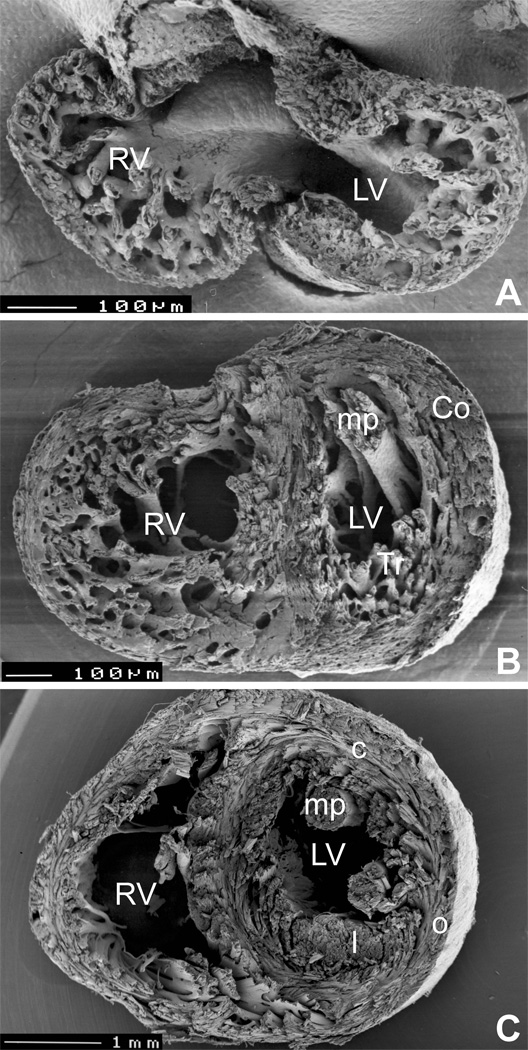
Figure 3.
Development of spiral architecture in the compact layer. A, pre-compaction stage (E13.5 embryonic mouse heart in transverse section) showing thin, avascular compact layer with circumferential arrangement of myocytes. B, post-compaction, vascularized compact layer at E15.5 shows spiral arrangement in both compact myocardium (Co) and trabeculae (Tr). C, in the adult mouse heart, the typical three-layered structure with inner predominantly longitudinal (l), middle circular (c) and outer oblique (o) orientation can be seen in the left ventricle (although recent quantitative studies show that the change in orientation occurs in continuum 29). LV, left ventricle, RV, right ventricle, mp, papillary muscles. Ventricular midportion slabs, viewed from the apex towards the outflow. The top two specimens were prepared by late Dr. Si Minh Pham at University of Lausanne.

