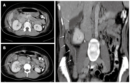
Figure 2.
A 31-year-old woman presenting with right hypochondrial pain and a clinical diagnosis of pelvic inflammatory disease and right pyelonephritis. A: Contrast-enhanced CT scan showing fluid collection (arrow) in the subhepatic region, extending anteriorly to the gallbladder fossa with inflammatory stranding; B: Note the air fluid level in the collection adjacent to the right kidney; C: Coronal reconstruction showing the long thickened and inflamed appendix (short arrows) reaching the subhepatic region, and the subhepatic collection (arrow) is seen extending more cranially.

