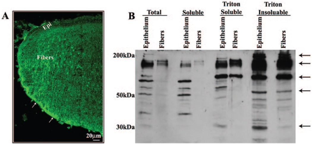
FIGURE 3.
Determination of spatial and subcellular distribution of ponsin in the mouse lens. (A) The distribution of ponsin in the lens epithelium and fiber cells was determined by labeling cryosections derived from 1-day-old mouse lenses with polyclonal ponsin antibody in conjunction with FITC-conjugated secondary antibody, followed by confocal image analysis. Arrows: an intense and somewhat punctate localization of ponsin to the posterior tips of the lens fibers and along the lateral membrane of the fiber cells. (B) The distribution profile of ponsin protein in different fractions of the lens was determined by subjecting epithelial and fiber cell protein fractions (40 µg/lane) derived from the adult mouse lens (total, soluble, and Triton-soluble and -insoluble fractions) to immunoblot analysis with polyclonal ponsin antibody. Multiple immunopositive ponsin-specific protein bands ranging in size from 30 to >150 kDa were identified in different fractions of the lens epithelium and fiber mass.

