
Fig. 1. Generation and characterization of keratinocyte strains expressing the HPV-16 E6 and E7 genes.
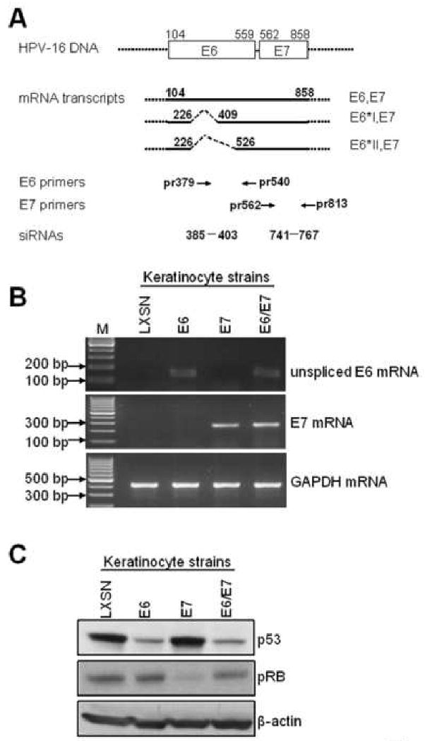
(A): Diagram of HPV-16 E6 and E7 mRNA expression and locations of primers and siRNA targets used in this study. The E6 and E7 open reading frames (ORFs) are shown as open boxes, with the nucleotide positions of the first nucleotide of the start codons and the last nucleotide of the stop codons indicated. The dotted lines flanking the open boxes and the heavy lines represent vector sequences. The heavy lines below the open boxes represent the E6/E7 bicistronic transcripts with alternative splicing sites depicted as dotted lines. The numbers above the full length E6E7 transcript are the nucleotide positions of the first nucleotide of the start codon of E6 and the last nucleotide of the stop codon of E7 in the HPV16 genome. The numbers above the E6*IE7 and E6*IIE7 transcripts are the positions of either 5′ or 3′ splicing sites. The primers used in this study are depicted below the transcript lines as arrows and numbers showing the locations of primers in the genome. The siRNA targets are also depicted as dashes and numbers showing the start and end nucleotides of the targets. (B): confirmation of E6, E7 mRNA expression. Primary human foreskin keratinocytes (HFK) were transduced with pLXSN expressing HPV-16 E6, E7, E6E7 or empty vector as described. Total cellular RNA was isolated from the transduced HFK cell strains and RT-PCR was performed with sets of HPV16 unspliced E6 and E7 specific primers described in Material and Methods, GAPDH mRNA was detected as an internal control. PCR products were analyzed with 2% agarose gel. (C): p53 and pRb proteins. To verify the expression of the E6 and E7 proteins, we screened the above stable cell lines by Western blotting for expression of p53 and pRb, respectively. Western blotting for β-actin also was used as a loading control. As anticipated, a decreased p53 protein was observed in E6 and E6E7 expressing cells, and decreased pRb was only observed in E7-expressing cells.