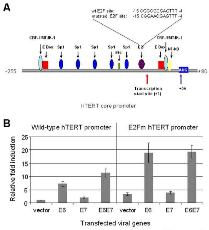
Fig. 6. E7 induces the hTERT promoter through an intact E2F site.

(A) Diagram of the hTERT core promoter. Binding sites for transcription factors are indicated as well as the transcription and translation start sites. The sequences and positions of wt and mutated E2F are also depicted. (B) E7 augments E6-induced hTERT promoter activity and this augmentation requires an intact E2F binding site. The wild-type hTERT core promoter (pGL3B-hTERT) was mutated at the E2F binding site using an overlapping PCR protocol described in Materials and Methods. Keratinocytes were transfected with either wt hTERT core promoter (pGL3B-hTERT) or the E2Fm mutant (pGL3B-hTERT-E2Fm) and either E6, E7 or both. The pRL-CMV R. reniformis reporter plasmid was also transfected into the cells to standardize for transfection efficiency. Relative fold activation reflects the normalized luciferase activity induced by E6 and E7 compared to the normalized activity of vector control. The value of pGL3B-hTERT activity with empty vector was set to 1. Error bars show the standard deviation for at least three independent experiments. The mutated E2F binding site in the core hTERT promoter led to an increased basal activity of the promoter and abrogated E7 induction and augmentation of the promoter. Mutation of the E2F binding site did not affect E6 induction of the promoter compared to vector.
