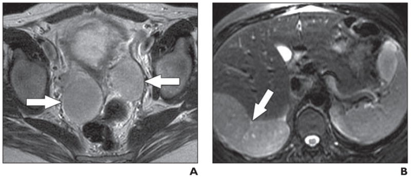
Fig. 12. 34-year-old pregnant woman.

A, Axial RARE T2-weighted MR image obtained at 23 weeks’ gestation shows bilateral solid adnexal masses (arrows).
B, Axial RARE T2-weighted MR image through upper abdomen shows large tumor deposit (arrow) abutting liver. Appearances are considered indicative of malignancy. Cesarean hysterectomy and bilateral salpingo-oophorectomy were performed at 28 weeks’ gestation because of progression of subphrenic tumor with diaphragmatic irritation. Pathology results showed benign metastasizing leiomyoma. Masses spontaneously regressed after surgery and patient remains free of disease 3 years after surgery.
