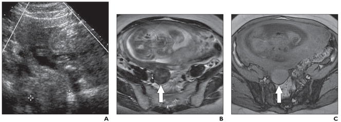
Fig. 2. 34-year-old pregnant woman who presented with lower abdominal pain.
A, Transvaginal sonogram obtained at 25 weeks’ gestation shows solid 3.8-cm mass (between calipers) thought to be of right adnexal origin.
B, Axial RARE T2-weighted image shows mass (arrow) arises from uterus and is of low T2 signal intensity; also, note “claw sign,” similar to Figure 1. Findings are those of exophytic uterine leiomyoma.
C, Axial spoiled gradient-echo T1-weighted MR image shows exophytic uterine leiomyoma (arrow) is of increased T1 signal intensity; this finding indicates red degeneration (i.e., spontaneous hemorrhagic infarction).

