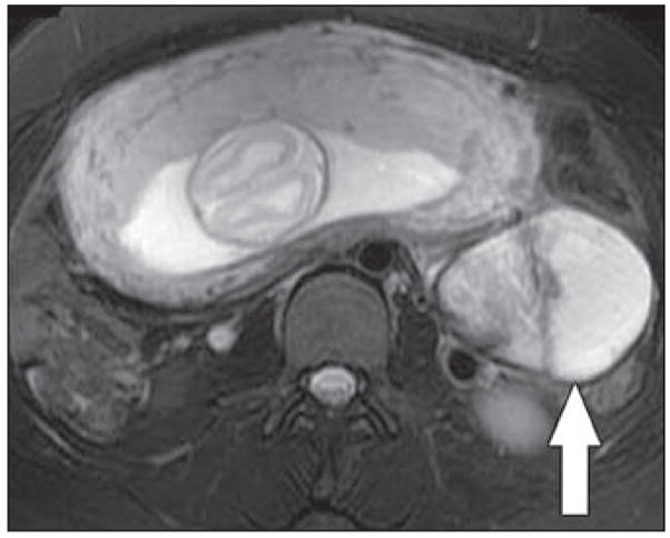
Fig. 4.

Axial RARE T2-weighted image with fat saturation obtained at 24 weeks’ gestation in 29-year-old woman shows complex mixed solid and cystic left adnexal mass (arrow). No increased signal was seen on T1-weighted images (not shown). MRI appearances are nonspecific, although diagnostic considerations include cystic malignancy. Mass was resected and found to be benign hemorrhagic cyst.
