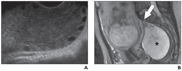
Fig. 8. 32-year-old woman with persistent right-sided pelvic pain.
A, Transvaginal sonogram obtained at 24 weeks’ gestation shows enlarged right ovary with preservation of peripheral follicles.
B, Sagittal RARE T2-weighted MR image shows right ovary (asterisk) is of markedly increased T2 signal intensity to degree that mass might be considered cystic if MRI findings had not been interpreted in conjunction with sonographic findings. Appearance is of massive ovarian edema. Cause of this condition is not well understood but may reflect chronic or subacute low-grade torsion. Beaklike pedicle (arrow) arising from superior aspect of ovary is compatible with this pathogenesis.

