Fig. 2.
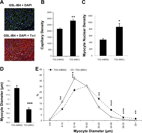
Increased capillary and myocyte nuclear densities after MSC administration. A: representative images of capillary and myocyte staining using FITC-labeled Griffonia simplicifolia isolectin B4 (GSL-IB4; green) and troponin I (TnI) antibody (red), respectively. 4′,6-Diamidino-2-phenylindole (DAPI; blue) was used for nuclear staining. B and C: computer analysis of capillary and myocyte nuclear densities (expressed as numbers/mm2). D: computer analysis of myocyte cross-sectional diameters. At least 15 fields of ×200 magnification and >350 myocytes were evaluated in each hamster. E: frequency histogram of diameters showing a greater number of smaller myocytes in the MSC-treated group (n = 4 animals/group). *P < 0.05 vs. control; **P < 0.01 vs. control; ***P < 0.001 vs. control.

