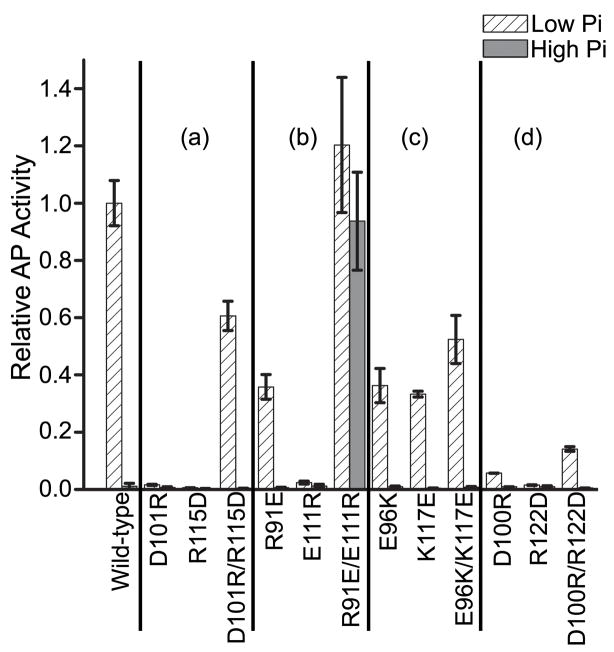
Fig. 8. Transcriptional activation of phoA by the single- and double-residue mutants that target the α4-β5-α5 surface.
Cells expressing the various mutants of PhoB were grown under low phosphate (inducing) and high phosphate (non-inducing) conditions and analyzed for AP activity as described in Materials and Methods. The “/” symbol is used to designate the double-residue mutants. For clarification, the results are grouped based on the salt bridge that they target. Group (a): D101…R115, group (b): R91…E111, group (c): E96…K117 and group (d): D100…R122. Each value represents a minimum of 3 replicates with error bars representing one standard deviation. The AP activity was normalized to the level attained by wild-type PhoB under low phosphate conditions.

