
Figure 4. The Intestinal Stem Cell Zone Replenishes the Adsorptive Villus.
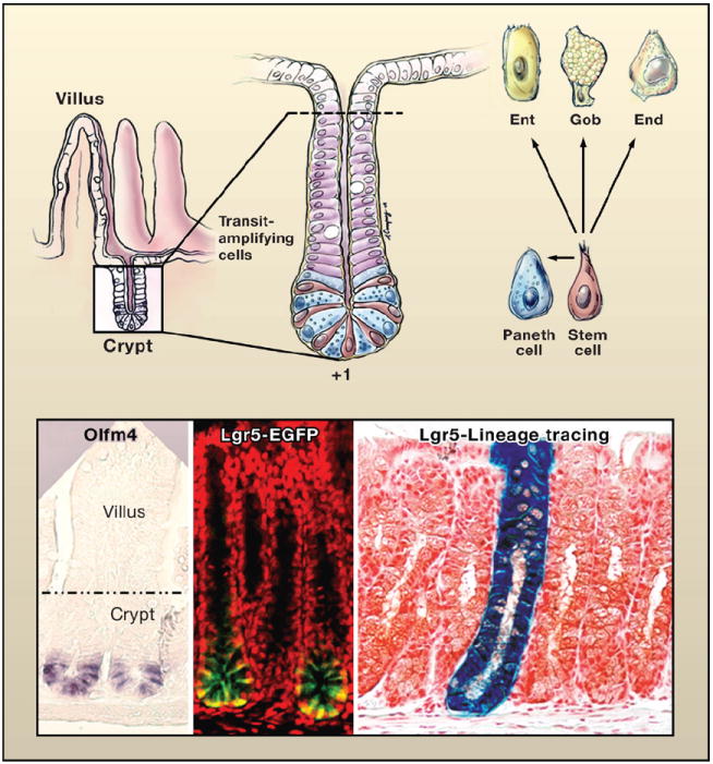
(Top left) Schematic of the crypt-villus architecture of the small intestine. (Top right) Magnified view of the crypt. At the base of the crypt is the putative stem cell zone (Bjerknes and Cheng, 1981; Cheng and Leblond, 1974). This zone contains undifferentiated cells sharing a similar ultrastructure that are interspersed with highly differentiated antibacterial Paneth cells. The undifferentiated columnar cells, purported to be stem cells, extend from a position +1 at the very base of the crypt to +4, marking the boundary between the Paneth cells and the transiently amplifying, rapidly dividing cells that give rise to the terminally differentiating enterocytes (Ent), goblet cells (Gob), and enteroendocrine cells (End) of the villus. Drawing by A. Canapary. (Lower panels) In situ hybridization with a probe for Olfm4, enriched in the stem cell zone (van der Flier et al., 2009). The expression of an Lgr5-EGFP-CreER transgene marks the stem cell zone (Barker et al., 2007). To the right is a lineage tracing of a bitransgenic mouse expressing Lgr5-EGFP-CreER and Rosa26-fl-stop-fl-LacZ. Cells in which the Lgr5 promoter is active are marked by EGFP expression and express tamoxifen-regulatable Cre recombinase. Following activation of Cre, floxed sequences recombine, excising the stop codon and activating LacZ. With time, the entire crypt-villus structure displays β-galactosidase expressing progeny, revealing that individual Lgr5-positive stem cells can regenerate the entire niche (Barker et al., 2007). Images reprinted by permission from Macmillan Publishers Ltd: Nature (Barker et al., 2007), copyright 2007.