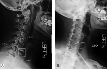
FIGURE 2.
(A) Conventional radiograph (lateral view) portraying an increased lordotic curvature and (B) with cervical spine positioned in neutral, revealing disc degeneration primarily at C6–C7 and C7–T1 (large arrows) and secondarily at C4–C5 (small arrows). The presence of osteophytes at C6–C7 are magnified in (A) and demarcated by the arrow in (B).

