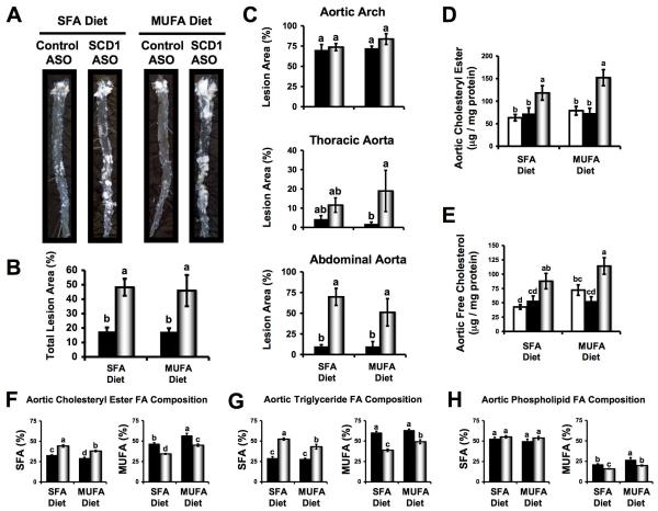
Figure 3.
SCD1 inhibition promotes atherosclerosis in LDLr-/-Apob100/100 mice. Starting at six weeks of age, mice were fed diets enriched in 0.1% (w/w) cholesterol and either saturated fatty acids (SFA) or monounsaturated fatty acids (MUFA) for 20 weeks in conjunction with biweekly injections (25 mg/kg) of either saline □, a non-targeting control ASO ■, or SCD1 ASO . (A) Representative photographs after en face preparation of aortae. (B) En face morphometric analysis of total aortic lesion area. (C) En face morphometric analysis of regional (aortic arch, thoracic aorta, and abdominal aorta) differences in atherosclerosis. Data shown in panels (B) and (C) represent the mean ± SEM from 6 mice per group. GLC analysis of aortic cholesteryl ester (D) and free cholesterol (E) was determined after morphometric analysis. Data shown in panels (D) and (E) represents the mean ± SEM from 8-15 mice per group. Fatty acid (FA) composition (% of total FA that was SFA or MUFA) of aortic cholesteryl esters (F), triglycerides (G), and phospholipids (H) was determined from whole aortic lipid extracts. Data shown in panels (F), (G), and (H) represents the mean ± SEM from 5 mice per group. Values not sharing a common superscript differ significantly (p<0.05).

