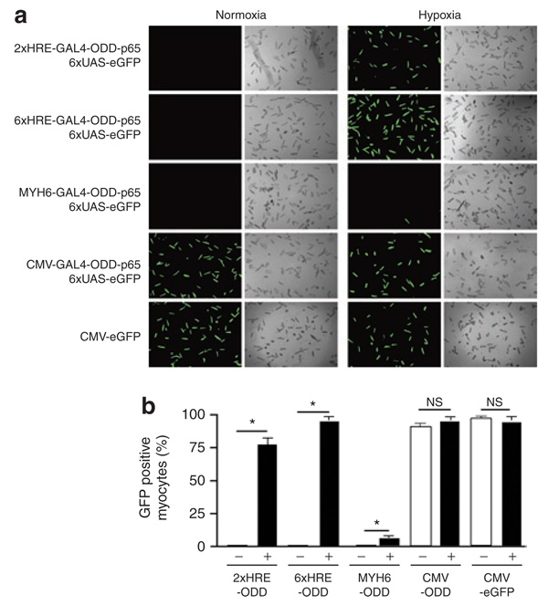
Figure 3. Hypoxia-mediated amplification of enhanced green fluorescent protein (eGFP) expression by double oxygen–sensing vector system.
Rat cardiac myocytes were transduced with identical sensor vectors as in Figure 1, containing unique promoters: 2xHRE-mp, 6xHRE-mp, Myh6, and cytomegalovirus (CMV) fused with GAL4- ODD-p65, and the 6UAS-eGFP effector virus was used as the reporter. (a) At 48 hours after transduction, we scored eGFP positive myocytes for each pair of viruses. (b) The percentage of GFP-positive myocytes within a cover slip was estimated by dividing GFP-positive by total myocytes. The CMV-eGFP virus served as a positive control for adenoviral transduction efficiency (a,b). In each graph, “+” is hypoxic and “−” is normoxic. Values are mean ± SEM. HRE, hypoxia-responsive element; NS, not significant; ODD, oxygen-dependent degradation domain.

