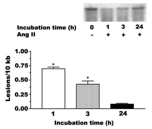
Figure 1).
Mitochondrial DNA damage mediated by angiotensin II (Ang II). Neonatal cardiomyocytes were incubated for various periods of time with medium containing 1 nM Ang II. High-molecular-weight DNA was isolated and digested to completion with BamHII. The samples were exposed to 0.1 M NaOH before electrophoresis on a 0.6% agarose gel. Shown is a representative Southern blot of the prominent 10 kb band of mitochondrial DNA. The reduction in the intensity of the 10 kb band is indicative of DNA fragmentation. The data are also expressed as the number of lesions per 10 kb. Although not shown, control samples contained no apparent lesion. Values shown represent the mean ± SEM of three to five preparations. *A significant difference between the Ang II-treated and control cells (P<0.05)

