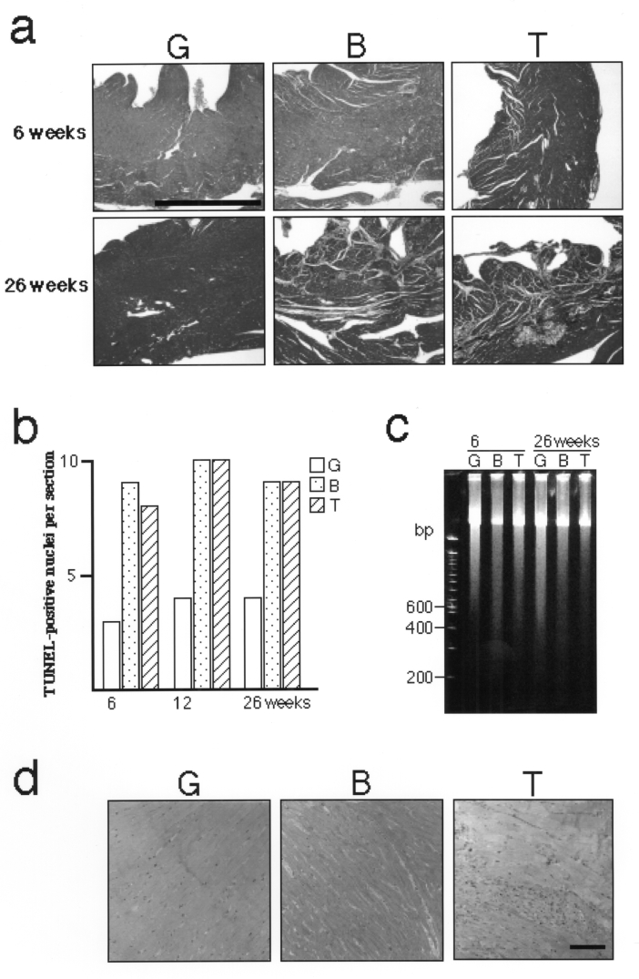
Figure 2).
Pathogenetic investigation of TO-2 hamsters. a Masson’s trichrome staining. Note marked fibrosis in the TO-2 hamster (bar = 1 mm). b The number of terminal deoxynucleotidyl transferase-mediated dUTP-biotin end-labelling (TUNEL)-positive nuclei per mid-line section of the hamster ventricle. No significant difference was observed between BIO14.6 and TO-2 hamsters. c Electrophoresis of genomic DNA from the left ventricle. No significant DNA ladder indicative of apoptosis was observed in BIO14.6 or TO-2 hamsters. d Toluidine blue staining of the left ventricle. Note the marked infiltrate of inflammatory cells in the TO-2 hamster (bar = 100 μm)

