
Figure 7.
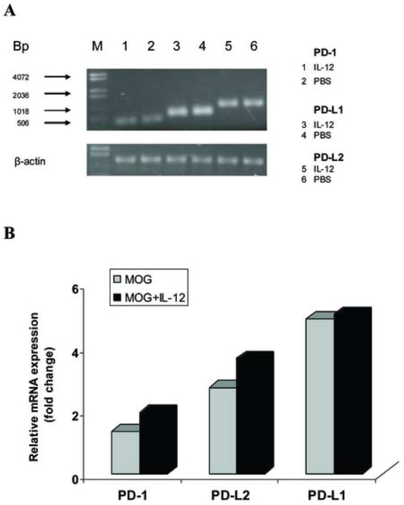
IL-12 treatment does not induce PD-L1 gene expression in MOG-specific T cells from IFN-γ-deficient mice in vitro. MOG-specific T cells isolated from inguinal and popliteal lymph nodes were prepared on day 10 p.i. from IFN-γ-deficient mice immunized with 300 μg MOG35-55 peptide. Total RNA was extracted from MOG-specific T cells cultured with MOG35-55 stimulation alone at a final concentration of 25 μg/ml (MOG), or cultured with MOG stimulation and co-cultured with 20 ng/ml rmIL-12 (MOG + IL-12). 5 μg RNA was used to generate cDNA. Quantitative RT-PCR was performed. β-actin was amplified in each sample as endogenous control. Each sample is triplicate. Quantization of gene expression was reported as the fold difference relative to the housekeeping gene. Electrophoresis of final products of PCR of PD-1, PD-L1 and PD-L2 gene and β-actin as endogenous control in 2.5% agarose-TBE gel is shown (A). There is no significant difference in PD-1, PD-L1 and PD-L2 gene expression between the “MOG” group and the “MOG + IL-12” group (p>0.05) (B). One representative experiment of three is shown.