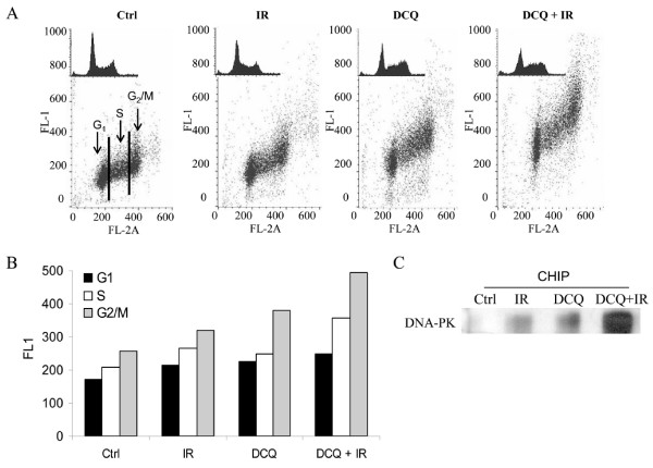
Figure 3.
Phosphorylation of ATM and activation of DNA-PK by DCQ ± IR in EMT-6 cells at 2 h post-treatment. A. EMT-6 cells were treated with 10 μM DCQ, 10 Gy IR, or combination treatments, fixed and subjected to immunocytochemical detection of ATM phosphorylated on Ser1981, and stained with PI to detect at the same time p-ATM in each phase of the cell cycle. B. The mean of the FL-1 intensity (reflecting the level of p-ATM expression) at each phase of the cell cycle are plotted. C. Anti-DNA-PK was immunoprecipitated with DNA from lysates of 106 EMT-6 cells treated with 10 μM DCQ, 10 Gy IR, or combination treatments using CHIP assay. The immunoprecipitate was resolved on a 5% gel by electrophoresis, transferred to nitrocellulose and probed with anti-DNA-PK. The bands were quantified using LabWorks 4.0 software. CHIP: chromatin immunoprecipitation.

